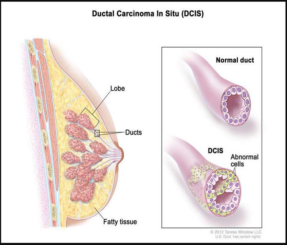
Ductal carcinoma in situ, or DCIS, is the earliest stage of breast cancer and is confined to the lining of the milk duct, the tube that carries breast milk from where it is made in the lobules to the nipple. The cancer is non-invasive, meaning it is “in its original place,” and has not spread beyond the milk duct into the surrounding breast tissue.
DCIS is categorized as a Stage 0 breast cancer.
According to the National Cancer Institute, about 80% of DCIS cases have no signs or symptoms of cancer and are found during a routine screening mammogram. The most common presentation is abnormal calcifications on imaging.
For the remaining 20% of cases, some of the signs or symptoms of breast cancer include:
- Lumps or bumps that don't go away
- Nipple discharge (clear or bloody)
- Dimpling of the skin
- Nipple retraction (pulling in) or inversion
- Nipple rash or itching
- Redness or swelling
- Changes to the skin's texture
- Breast or nipple pain or discomfort
- Swelling to the axillary area (armpit)
Any changes to your breast or axillary area should be reported to your physician as soon as possible for further evaluation and workup.
Imaging
Imaging tests such as a mammogram, ultrasound, and breast MRI take pictures of the inner part of your breast(s) so that a radiologist, a doctor that specializes in reading images, can take a closer look at your body.
-
Mammography uses X-rays to create images of your breasts
-
Screening Mammograms are done when there are no symptoms
- Example: annual mammograms
- Diagnostic mammograms are done when there is an area of concern
- 3D digital imaging or breast tomosynthesis is an advanced form of imaging that offers high-quality images from various angles, resulting in more accurate results vs. 2D imaging
-
Screening Mammograms are done when there are no symptoms
-
Ultrasound uses sound waves to create images of the breast tissue
- This procedure is often used in combination with a mammogram to evaluate areas of concern
- The procedure is painless
-
Breast MRI is indicated in some individuals.
- The MRI is radiation free and it creates a 3-D image of the breasts
- Helps with staging and pre-surgery planning
- The procedure uses an IV contrast which is injected intravenously into the arm before the test. This contrast lights up during the exam, giving the doctor a better look at the breasts and surrounding areas
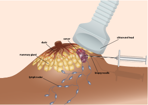
Breast Biopsy
A breast biopsy is the only diagnostic procedure that can definitively confirm or rule out a breast cancer diagnosis.
When a suspicious mass or an area of concern is seen on a mammogram or ultrasound, your doctor will order a breast biopsy to have a sample of the tissue examined to check for the presence of cancer cells.
After the tissue from the breast biopsy is removed, it is sent to a lab where a pathologist, a doctor that specializes in analyzing body tissue, examines the cells under a microscope and determines a diagnosis. The pathologist will interpret the tissue samples and prepare a report to share with the healthcare team.
The pathology report will contain some of the following information about the diagnosis:
- Procedure performed (type of biopsy)
- Specimen location (breast, axilla, lymph node(s), etc.)
- Histology type (ductal, lobular, phyllodes, etc.)
-
Grade
- Low - Grade I – cells look most like normal breast cells and are usually slow growing
- Intermediate - Grade II – cells look less like normal breast cells and are faster growing
- High - Grade III – cells look the least like normal breast cells and are usually fast-growing
-
Prognostics/receptors/biomarkers
- The pathologist will look for estrogen receptors (ER) and progesteronereceptors (PR) on the breast cancer cells to determine the tumormarker status
-
About half of all DCIS are ER and/or PR positive
-
If breast cancer cells have ER and/or PR, they are called ER and/orPR positive
- Tumors that are ER and/or PR positive are more likely torespond to hormone therapy
-
If breast cancer cells do not have ER and/or PR, they are called ER and/or PR negative
- Tumors that are ER and/or PR negative are less likely to respondto hormone therapy
-
If breast cancer cells have ER and/or PR, they are called ER and/orPR positive
Surgery
Surgery is one of the common treatments that you can expect after being diagnosed with DCIS. When speaking with your surgeon, you will learn about the different surgical options that are recommended for you.
These recommendations will be based on many factors:
- Stage, tumor size, and the location of your tumor
- Lymph node involvement (not likely with DCIS)
- The size of your breasts
- Your risk of cancer recurrence
- Age and personal preferences
Two of the main types of breast cancer surgeries are called partial mastectomy (also called lumpectomy) and mastectomy, which are performed by a surgeon. If you choose to have a mastectomy, you will have the option to have reconstruction surgery, which is performed by a plastic surgeon.
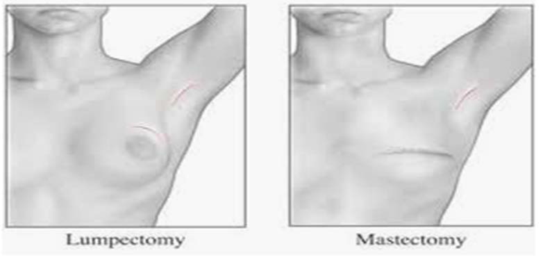
Partial Mastectomy or Lumpectomy
A partial mastectomy, also called lumpectomy, segmental mastectomy, or quadrantectomy, is considered breast-conserving-surgery (BCS) and is the most common type of surgery for women with DCIS.
During a partial mastectomy, the surgeon removes the area of disease and a margin of surrounding healthy breast tissue. The goal of a partial mastectomy is to remove the tumor with negative margins and to preserve the appearance and shape of the breast.
Mastectomy
A mastectomy is a surgical procedure involving the complete removal of the breast.
Mastectomies can be categorized into several different types:
- Nipple-sparing mastectomy – the breast tissue is removed preserving the overlying skin, nipple, and areola. This technique is utilized in patients pursuing breast reconstruction. Candidacy for nipple preservation is influenced by tumor factors and patient factors.
- Skin-sparing mastectomy – the breast is removed preserving the overlying skin but removing the nipple and areola. This technique is utilized in patients pursuing breast reconstruction.
- Total mastectomy - the breast is removed along with overlying breast skin, nipple, and areola. A total mastectomy leaves a flat contour to the chest following surgery.

Most insurance companies will cover the cost of mastectomy bras, breast forms, camisoles, and prostheses. Ask your surgeon to write a prescription for these medically necessary supplies.
Breast Reconstruction Surgery
Breast reconstruction surgery is a treatment option for you to consider if you choose a mastectomy. This procedure is done by a plastic surgeon, a physician who specializes in reconstructing the breast mound after a mastectomy.
The goal of breast reconstruction surgery is to restore your breast(s) appearance and help you feel better about your body and the way you look in clothing after having a mastectomy. The plastic surgeon will talk to you about your treatment options, all the while, keeping in mind your lifestyle and personal preferences.
The two main types of reconstructive surgery are breast implants and autologous reconstruction (using your own tissue to rebuild the breast mound). Keep in mind that every woman’s circumstance is different and recovery times post-surgery can vary greatly based on the type of surgery you choose.
Does Surgery Choice Affect Recurrence Rates?
Even though DCIS is stage 0 breast cancer, that doesn’t make surgical decisions any easier. A common dilemma that women face after a breast cancer diagnosis is deciding on the type of surgery to have in order to give them the best chance that the cancer will not recur.
Regardless of your surgery choice, long-term survival rates are the same, and local recurrence rates remain about the same when the standard of care is followed and are influenced by the following tumor characteristics such as the grade, size of the tumor, and margin status.
Partial Mastectomy or Lumpectomy
- Women who have a partial mastectomy followed by radiation therapy have about a 15% recurrence rate
- Women who have a partial mastectomy with no other treatment have about a 25 to 30% recurrence rate
Mastectomy
- Women who have a mastectomy will have a slightly lower risk of breast cancer recurrence compared to women who have a partial mastectomy with radiation
- Survival rates for women that have a mastectomy are the same as for women who have a partial mastectomy with radiation
Nationally, the most common type of treatment for women with DCIS is a partial mastectomy followed by radiation therapy and endocrine therapy if the tumor is estrogen receptor-positive.
Your healthcare team can help create a plan that is specific for you based on your medical history, tumor characteristics, and personal feelings.

After receiving a DCIS diagnosis, you can expect to see a medical oncologist at some point in your treatment plan (typically after surgery). The medical oncologist has extensive knowledge in diagnosing and treating cancer and pays special attention to the pathology report(s) from your breast biopsy(s) and surgical pathology. The information in the pathology report helps the physician determine the best treatment option for the patient.
In addition, the medical oncologist will discuss the risk of your cancer coming back after you have completed treatment.
Sometimes, additional testing (such as imaging, tissue sampling or genetic testing) will be needed to gather more information about your disease before a treatment plan can be established.
Hormone Blocker or Endocrine Therapy
A hormone blocker or endocrine therapy is prescribed for hormone receptor positive tumors and is a type of “systemic treatment,” meaning it affects the entire body, not just the breast.
The goal of a hormone blocker is to kill cancer cells and reduce the risk of recurrence:
- Cancer cells have receptors (proteins) which attach to estrogen and progesterone and the hormones “feed” the cells, making them grow out of control and spread
- Hormone-blockers can either stop the hormones from attaching to the receptors or stop estrogen production
The medication comes in pill form and is taken daily for five years. However, some patients may benefit from taking the medication for 10 years or more, depending on the circumstances. A medical oncologist will help determine which medication is best for you based on your medical history, stage of disease and menopausal status.
Hormone-blockers are typically prescribed after local treatment (surgery or radiation therapy) has been completed. In some circumstances, a physician may prescribe the medication sooner if surgery is delayed or the disease has spread to other organs (not likely with DCIS). Patients who undergo a bilateral mastectomy may be able to avoid endocrine therapy post-surgery. Talk to your medical oncologist to see what option is best for you.
If your pathology report indicates that your cancer is hormone receptor positive, your medical oncologist will determine which medication is most beneficial to you based on your medical history and menopausal status.
Two types of hormone-blockers are:
- Tamoxifen - oral medication that blocks estrogen from attaching to cancer cells. Tamoxifen is typically used for women that are pre-menopausal but can also be used for post-menopausal women.
-
Aromatase Inhibitors (AI) - oral medications that stop estrogen production in the adrenal glands. Aromatase inhibitors are typically used for women that are post-menopausal, but also can be used for women who are pre-menopausal and are taking an ovarian suppression medication.
- Three types of AI's include:
- Arimidex (anastrozole)
- Aromasin (exemestane)
- Femara (letrozole)
There are medications that can block the anti-cancer effectiveness of hormone-blockers so talk to your medical oncologist about what medications to avoid while on treatment.
Side Effects of Hormone Blockers
Not everyone experiences side effects of hormone blockers, but like any medication, they have the potential risk of side effects.
Knowing that the hormone-blockers can reduce the risk of the breast cancer coming back often helps women stay with the medication despite the side effects.
Some of the potential side-effects of Tamoxifen:
-
Menopausal Symptoms
- Hot flashes
- Vaginal dryness or discharge
- Abnormal vaginal bleeding or loss of menstrual cycles
- Loss of libido
- Frequent UTI's
-
GI Symptoms
- Nausea
- Constipation
- Weight gain
- Leg swelling
- Mood swings
- Depressing
- Headaches
- Brain fogginess
- Fatique
- Weakness
- Bone pain
- Dry skin
- Hair loss
-
Beneficial side effects
- Helps stop bone loss after menopause
- Lower cholesterol levels
In rare instances, Tamoxifen can cause uterine cancer, blood clots and stroke.
Some of the potential side-effects of aromatase inhibitors:
- Bone, joint or muscle achiness/bone thinning/broken bones
- Brain fogginess/mood disturbances
- Insomnia
- Heart problems/peripheral edema/high blood pressure
- Hot flashes
- Stomach upset/nausea/vomiting
- Decreased energy
- Headache
- Elevate cholesterol levels
Not all side effects are listed above. It is recommended that you carefully review the package insert to educate yourself and inform your healthcare provider if you experience any unusual symptoms.
In some circumstances, women may go through a “menopause transition” while on Tamoxifen and become post-menopausal. These women may switch to an AI after 2-3 years.
While studies have shown that Tamoxifen is associated with preservation of bone mineral density, AI’s can have the opposite effect, often contributing to net bone loss.
Therefore, the following is recommended while on an AI:
- Baseline bone density test and repeated every 2 years
- Exercise regularly to increase bone strength
-
Adequate amount of calcium in your diet
- Dairy products, almonds, broccoli, kale, salmon, sardines, tofu
-
Adequate amount of Vit D in your diet
- Fortified dairy products/tuna/egg yolks/liver
- No smoking
- Limit drinking alcohol
-
Calcium and Vit D supplements (pharmaceutical grade or USP)
- Discuss the recommended dose with your physician

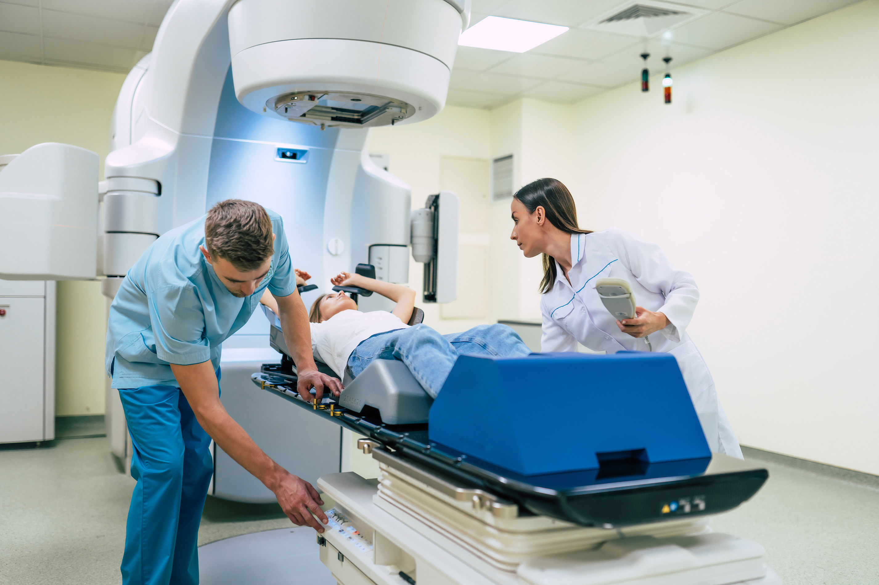
Radiation therapy is a local cancer treatment that uses high energy x-rays to kill cancer cells and shrink tumors. If you are a candidate for this type of treatment, you will be referred to a radiation oncologist, a physician who treats cancer with radiation therapy.
The radiation oncologist will likely recommend treatment based on the following factors:
- After a partial mastectomy
- Helps lower the risk of the cancer coming back in the treated breast or in nearby lymph nodes
Radiation therapy is an outpatient procedure that typically starts after your surgical wound has healed (roughly 4 weeks). The radiation oncologist will set up a treatment plan specially designed for you through careful planning and mapping.
During this planning stage, you will be given special skin markings, which will aid in proper positioning for daily treatments. Radiation is given daily, Monday through Friday, for 3-6 weeks and each treatment takes only 10-15 minutes.
Some women have a family history of breast, ovarian, colon or pancreatic cancer and may be at a higher risk of developing a second cancer (in the same or opposite breast) or another type of cancer during their lifetime due to gene mutations in their DNA.
Your physician will review your heredity cancer risk at the time of your consultation and determine if you meet the guidelines for genetic testing.
There are a variety of sophisticated genetic tests available today that are performed by obtaining a sample of your blood or saliva. The test contains a panel of genes with each gene carrying a different risk for future cancers.
According to the American Cancer Society, the most common type of heredity breast cancer is found in the BRCA1 or BRCA2 gene. Women with one of these mutations are not only more likely to develop breast cancer in her lifetime, but also at a younger age. They are also at a higher risk to develop ovarian and other associated cancers in their lifetime.
If an inherited gene is found, treatment options can become more complex and the patient should consult with their healthcare team about the current guidelines of care. Keep in mind that not everyone who carries a gene mutation develops breast or ovarian cancer.
According to the American Cancer Society, there is no way to prevent a recurrence of cancer, but there are things that you can do to decrease your chances of a recurrence:
- Get regular screening
-
Stay active and maintain a healthy body mass index (BMI)
- Getting at least 5 hours of exercise per week can decrease recurrence ≥ 30%
-
Eat well-balanced meals
- Reduce red meat intake
- Increase daily fiber intake
- Avoid trans fats
- Limit or avoid simple sugars
- Anti-inflammatory diet (fruits/vegetables/omega-3 fatty acids, whole grains, lean protein, etc.)
- Consume fish several times weekly
- Consume at least five servings of fruits and vegetables daily
- Consume folate rich foods daily (legumes, leafy vegetables, asparagus, eggs, beets, citrus fruits, etc.)
- Limit or avoid alcohol
- Do not smoke
After cancer treatment is completed, your healthcare team will set-up a post-treatment follow-up or survivorship care plan with you. The goal of this plan is to provide patients with an individualized care plan that addresses follow-up visits as well as long-term side effects of cancer and its treatments.
Each team will have their own set of guidelines for follow-up care, so talk to them individually to establish a plan for your post-treatment needs.
Some of the services that can be expected during your follow-up care include:
-
Physician visits
- Each specialist has their own set of guidelines for follow-up care
- Emotional well-being check
-
Imaging studies
-
Varies based on the type of surgery and age of the patient
- Mammogram/Ultrasound/MRI/ Bone density test
-
Varies based on the type of surgery and age of the patient
-
Managing side-effects
- Medication education
- Ways to reduce side effects
- Physical therapy (if applicable)
-
Lifestyle modification recommendations
- Eat a healthy diet and exercise regularly
- No smoking
- Limit or avoid alcohol
- Avoid environmental toxins
- Get enough sleep
- Manage stress
- Surround yourself with supportive people
National Cancer Institute:
https://www.cdc.gov/cancer/breast/basic_info/symptoms .htm
American Cancer Society:
https://www.cancer.org/research/cancer-facts-statistics/breast-cancer-facts-figures.html
https://www.cancer.org/cancer/breast-cancer/risk-and-prevention/can-i-lower-my-risk.html
https://www.cancer.org/cancer/breast-cancer/risk-and-prevention/breast-cancer-risk-factors-you-cannot-change.html
https://www.cancer.org/cancer/breast-cancer/treatment/surgery-for-breast-cancer/mastectomy.html
https://www.cancer.org/cancer/breast-cancer/treatment/surgery-for-breast-cancer/breast-conserving-surgery-lumpectomy.html
https://www.breastcancer.org/symptoms/testing/types/mri/screening
Images:
https://www.cancer.gov/news-events/cancer-currents-blog/2015/dcis-low-risk
https://hlp.nucleushealth.com/lumpectomy-and-mastectomy-appearance-after-surgery/view-item?ItemID=10385